BPC-157 in cultured rat tendon fibroblasts: a published study summary
Summary
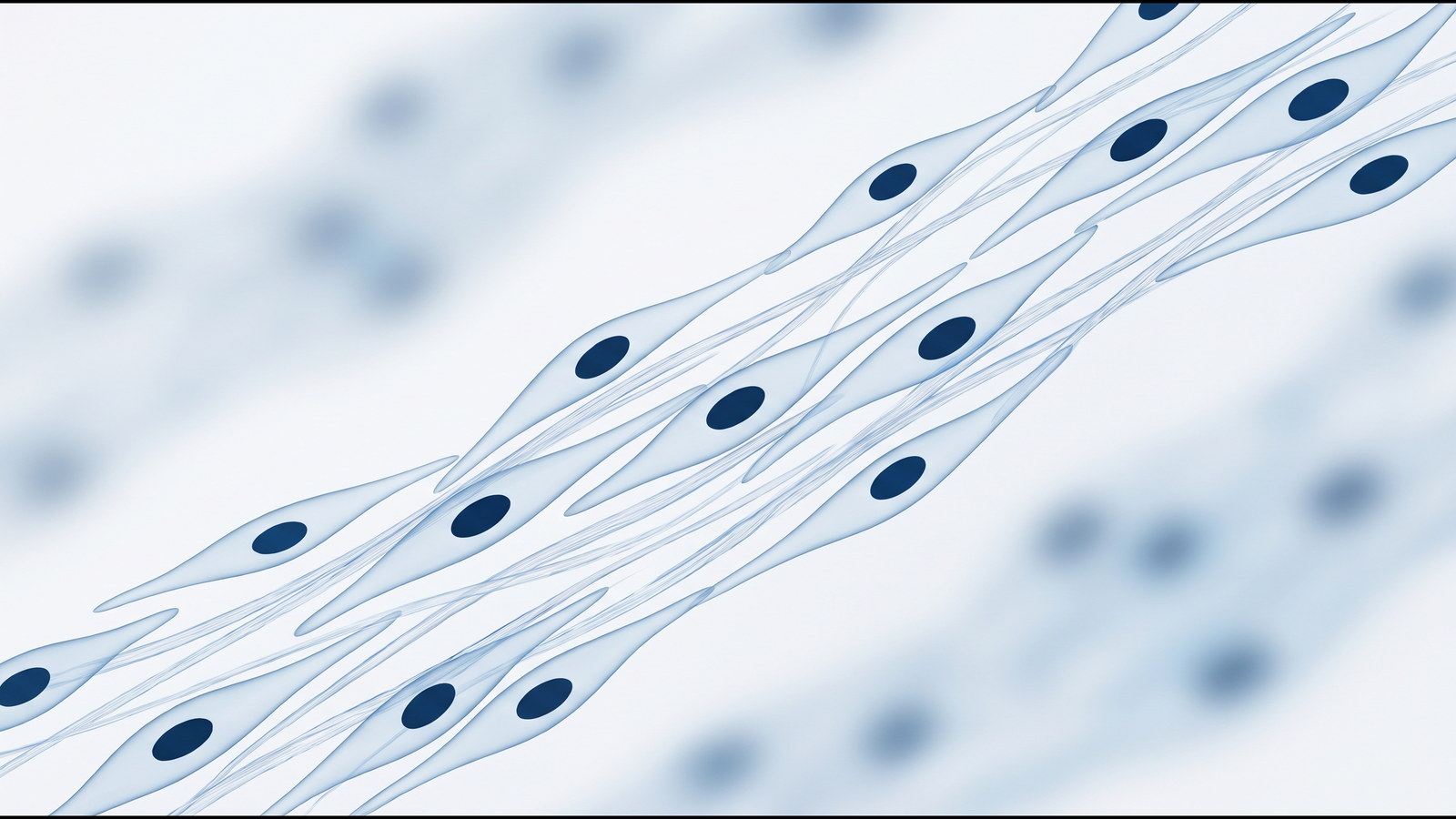
This summary describes a cell-culture study of the pentadecapeptide BPC-157 published in the Journal of Applied Physiology in 2011 by Chang and colleagues at Chang Gung University. The authors prepared tendon explants and primary tendon fibroblasts from rat Achilles tendon and cultured them with or without BPC-157 in the medium. The endpoints were fibroblast outgrowth from explants, cell proliferation by MTT assay, cell survival after hydrogen peroxide exposure, transwell migration, cell spreading, F-actin staining, and the phosphorylation state of focal adhesion kinase (FAK) and paxillin by western blot. No animal or human endpoints were part of this experiment.
Peptide
- Name
- BPC-157
- Sequence
- Gly-Glu-Pro-Pro-Pro-Gly-Lys-Pro-Ala-Asp-Asp-Ala-Gly-Leu-Val
- CAS
- 137525-51-0
- Formula
- C62H98N16O22, approximately 1419.5 g/mol
- Class
- Synthetic pentadecapeptide. The authors describe it as a partial sequence of a protein isolated from human gastric juice.
Study design
Model system: tendon explants and primary tendon fibroblasts obtained from rat Achilles tendon and maintained in standard culture.
Groups: explants and cell cultures with BPC-157 added to the culture medium, compared with cultures maintained in medium without the peptide. The abstract describes several of the responses as concentration dependent, so a series of peptide concentrations was applied to the cultures. The full paper gives the concentration range. Nothing was given to a living animal in this study; the peptide was only present in the culture medium.
Endpoints measured:
- Outgrowth of fibroblasts from tendon explants, observed in culture.
- Proliferation of cultured tendon fibroblasts by MTT assay.
- Cell survival under hydrogen peroxide exposure.
- Migration of fibroblasts through a transwell filter.
- Spreading of fibroblasts on culture dishes.
- F-actin formation by FITC-phalloidin staining.
- Protein expression and phosphorylation of FAK and paxillin by western blot.
Duration: the culture periods differ by assay and are stated in the full paper.
Reported findings
The authors reported that outgrowth of fibroblasts from tendon explants was accelerated in the cultures that contained BPC-157 compared with the cultures that did not.
Proliferation of cultured tendon fibroblasts, measured by MTT assay, was not directly affected by the peptide.
Survival of cells exposed to hydrogen peroxide was reported as significantly higher in the BPC-157 cultures than in the cultures without the peptide.
Migration of tendon fibroblasts across a transwell filter increased with peptide concentration. Spreading of fibroblasts on culture dishes was also reported to increase with concentration.
FITC-phalloidin staining showed F-actin formation in fibroblasts exposed to BPC-157.
Western blot analysis showed that phosphorylation of both FAK and paxillin increased with peptide concentration, while the total amounts of FAK and paxillin protein were unchanged.
The abstract reports these differences as statistically significant; the exact test statistics, p-values and fold changes are given in the tables and figures of the full paper and are not reproduced here. The authors interpreted the migration and spreading observations as likely to be mediated by activation of the FAK-paxillin pathway.
Limitations
The experiments were performed in a single laboratory on primary cells and explants from rats. Cell-culture responses do not establish what happens in a whole organism. Group sizes per assay are given only in the full paper. The FAK-paxillin interpretation rests on phosphorylation measurements and was not tested with pathway inhibitors in the abstract. No human data were reported.
References
- Chang CH, Tsai WC, Lin MS, Hsu YH, Pang JH. Journal of Applied Physiology. 2011;110(3):774-780. PMID 21030672. DOI 10.1152/japplphysiol.00945.2010.
- Hsieh MJ, Liu HT, Wang CN, Huang HY, Lin Y, Ko YS, Wang JS, Chang VH, Pang JS. Journal of Molecular Medicine. 2017;95(3):323-333. PMID 27847966. DOI 10.1007/s00109-016-1488-y.
- Staresinic M, Sebecic B, Patrlj L, et al. Journal of Orthopaedic Research. 2003;21(6):976-983. PMID 14554208. DOI 10.1016/S0736-0266(03)00110-4.
This summary describes published research for the information of qualified researchers. It is not a description of any product sold on this site, which are supplied for in-vitro laboratory research only and are not for human or veterinary use.