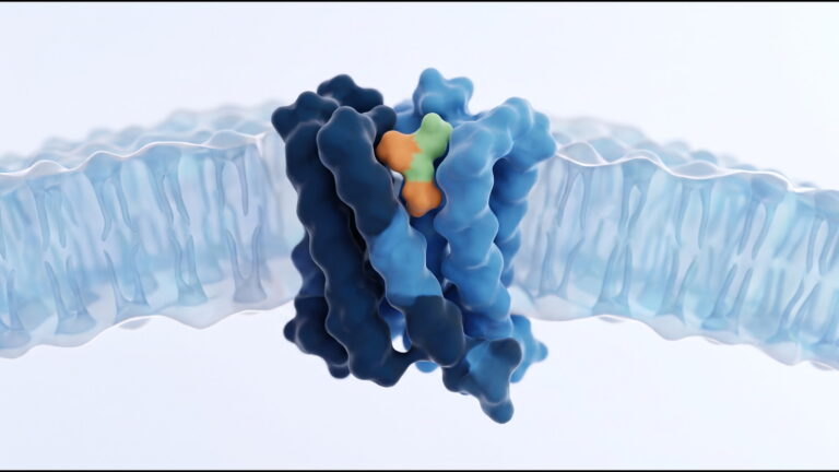
MOTS-c in cultured human cells: nuclear translocation and gene expression under nutrient restriction, a published study summary

Summary
This summary describes a 2018 study in Cell Metabolism by Kim, Son, Benayoun and Lee at the University of Southern California, together with the 2015 paper in the same journal by Lee and colleagues that first reported the peptide. MOTS-c is a 16-residue peptide encoded by a short open reading frame within the mitochondrial 12S ribosomal RNA gene. The 2018 work followed the subcellular location of MOTS-c in cultured cells placed under nutrient restriction and examined which nuclear genes changed expression and which transcription factors the peptide associated with. The 2015 work described the open reading frame and the cellular pathway affected in culture.
Peptide
- Name
- MOTS-c (mitochondrial open reading frame of the 12S rRNA type-c)
- Sequence
- Met-Arg-Trp-Gln-Glu-Met-Gly-Tyr-Ile-Phe-Tyr-Pro-Arg-Lys-Leu-Arg (MRWQEMGYIFYPRKLR)
- CAS
- 1627580-64-6
- Formula
- C101H152N28O22S2, approximately 2174.6 g/mol
- Class
- Mitochondrial-derived peptide encoded in mitochondrial DNA.
Study design
Model system (Kim 2018): cultured human cell lines, principally HEK293 cells, maintained in standard medium or placed under restriction of the principal carbohydrate in the medium to impose nutrient stress.
Procedures: subcellular location of MOTS-c was followed by immunofluorescence and cell fractionation, with mitochondrial, cytoplasmic and nuclear fractions blotted separately. Dependence on AMP-activated protein kinase (AMPK) was tested with inhibition and activation of that kinase. Gene expression was profiled by RNA sequencing. Association of MOTS-c with chromatin and with transcription factors was examined by chromatin immunoprecipitation and co-immunoprecipitation.
Endpoints measured:
- Nuclear versus cytoplasmic and mitochondrial location of MOTS-c under normal and restricted nutrient conditions.
- Dependence of translocation on AMPK.
- Sets of nuclear genes whose expression changed under nutrient restriction in a MOTS-c-dependent manner.
- Physical association with transcription factors that act on antioxidant response elements (ARE), including NFE2L2 (NRF2).
Model system (Lee 2015): a bioinformatic search of the mitochondrial genome for short open reading frames, followed by cell-culture experiments on the pathway affected by the synthetic peptide. The same paper also reports experiments in mice; those are outside the scope of this summary.
Reported findings
Kim and colleagues reported that MOTS-c translocated to the nucleus following nutrient restriction and that this translocation was dependent on AMPK. In the nucleus, MOTS-c was reported to regulate a broad range of genes in response to the restriction, including genes carrying antioxidant response elements, and to interact with ARE-regulating stress-responsive transcription factors such as NFE2L2/NRF2. The authors interpreted the findings as evidence that a peptide encoded in the mitochondrial genome can act on nuclear gene expression.
Lee and colleagues reported a short open reading frame within the mitochondrial 12S rRNA encoding the 16-amino-acid peptide. In cultured cells, the peptide’s actions were reported to inhibit the folate cycle and the de novo purine biosynthesis tethered to it, leading to activation of AMPK.
The gene lists, fold changes, immunofluorescence images and statistical values are in the full papers and are not reproduced here.
Limitations
The nuclear-translocation study was carried out in immortalised cell lines in one laboratory, and the senior author declares a consultancy and shareholding in a company working on mitochondrial-derived peptides. Subcellular localisation by immunofluorescence depends on antibody specificity, and nutrient restriction in culture is an artificial state whose relation to any physiological state is not established by the study. The mouse arm of the 2015 paper is not summarised here. No human data are summarised.
References
- Kim KH, Son JM, Benayoun BA, Lee C. Cell Metabolism. 2018;28(3):516-524.e7. PMID 29983246. DOI 10.1016/j.cmet.2018.06.008.
- Lee C, Zeng J, Drew BG, et al. Cell Metabolism. 2015;21(3):443-454. PMID 25738459. DOI 10.1016/j.cmet.2015.02.009.
- Mangalhara KC, Shadel GS. Cell Metabolism. 2018;28(3):330-331. PMID 30184481. DOI 10.1016/j.cmet.2018.08.017.
This summary describes published research for the information of qualified researchers. It is not a description of any product sold on this site, which are supplied for in-vitro laboratory research only and are not for human or veterinary use.