Epithalon (Ala-Glu-Asp-Gly): uptake of a FITC-labelled tetrapeptide by HeLa cells and in-vitro DNA binding, a published study summary
Summary
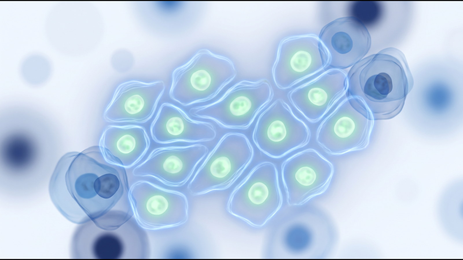
This summary describes a 2011 study in Biochemistry (Moscow) by Fedoreyeva, Kireev, Khavinson and Vanyushin, with a companion paper from the same group in the Bulletin of Experimental Biology and Medicine. The authors labelled the tetrapeptide Ala-Glu-Asp-Gly (epithalon) and two other short peptides with fluorescein isothiocyanate (FITC), incubated HeLa cells with them, and recorded where fluorescence appeared inside the cells. They then measured, in cell-free solution, how the unlabelled peptides quenched the fluorescence of carboxyfluorescein-labelled deoxyribooligonucleotides and of DNA-ethidium bromide complexes, and used Stern-Volmer constants to compare binding to different nucleotide sequences.
Peptide
- Name
- Epithalon (also written Epitalon)
- Sequence
- Ala-Glu-Asp-Gly (AEDG)
- CAS
- 307297-39-8
- Formula
- C14H22N4O9, approximately 390.3 g/mol
- Class
- Synthetic tetrapeptide.
Study design
Model system: HeLa cells in culture for the uptake experiments; cell-free fluorescence measurements for the binding experiments.
Materials: FITC-labelled epithalon (Ala-Glu-Asp-Gly), pinealon (Glu-Asp-Arg) and testagen (Lys-Glu-Asp-Gly) for cell uptake; unlabelled peptides, including bronchogen (Ala-Glu-Asp-Leu), for the binding experiments; 5,6-carboxyfluorescein-labelled single- and double-stranded deoxyribooligonucleotides of defined sequence, including sequences with and without cytosine methylation; DNA-ethidium bromide complexes.
Procedure: HeLa cells were incubated with each FITC-labelled peptide and examined by fluorescence microscopy. In the cell-free experiments, fluorescence of the labelled oligonucleotides or of the DNA-ethidium bromide complex was recorded as peptide was added, and Stern-Volmer quenching constants were calculated.
Endpoints measured:
- Location of fluorescence in HeLa cells (cytoplasm, nucleus, nucleolus).
- Stern-Volmer quenching constants for each peptide against each labelled oligonucleotide.
- Preference of each peptide for particular trinucleotide sequences and for methylated versus unmethylated cytosine.
Companion paper (Khavinson 2011): hydrolysis of lambda phage DNA by the eukaryotic endonucleases WEN1 and WEN2 was measured in the presence of short peptides of two to four residues, with and without histone H1.
Reported findings
The authors reported marked fluorescence in the cytoplasm, nucleus and nucleolus of HeLa cells after incubation with each of the FITC-labelled peptides, including epithalon.
In the cell-free experiments, the Stern-Volmer constants for quenching of the labelled single- and double-stranded oligonucleotides differed according to the primary structure of the peptide. The authors interpreted this as sequence-dependent binding. Epithalon, pinealon and bronchogen were reported to bind preferentially to oligonucleotides containing a CNG sequence. Epithalon, testagen and pinealon were reported to prefer CAG-containing sequences, while bronchogen preferred CTG. The peptides were reported to distinguish the cytosine methylation status of the oligonucleotides.
In the companion paper, short peptides were reported to inhibit or stimulate hydrolysis of lambda phage DNA by WEN1 and WEN2 depending on the DNA methylation status, to change the set of sites hydrolysed in double-stranded substrates, and to show different effects depending on their primary structure, with histone H1 modulating the effect.
The numerical Stern-Volmer constants and the microscopy images are in the full papers and are not reproduced here.
Limitations
Both papers come from the same group and a single laboratory. The uptake observations are qualitative fluorescence microscopy of a transformed cell line; a FITC label changes the size and charge of a tetrapeptide, so uptake of the labelled peptide may differ from that of the unlabelled peptide. The binding measurements are cell-free and rest on fluorescence quenching, which reports proximity rather than a defined complex. No animal or human data were reported.
References
- Fedoreyeva LI, Kireev II, Khavinson VKh, Vanyushin BF. Biochemistry (Moscow). 2011;76(11):1210-1219. PMID 22117547. DOI 10.1134/S0006297911110022.
- Khavinson VKh, Fedoreyeva LI, Vanyushin BF. Bulletin of Experimental Biology and Medicine. 2011;151(1):66-70. PMID 22442805. DOI 10.1007/s10517-011-1261-8.
- Khavinson VKh, Bondarev IE, Butyugov AA. Bulletin of Experimental Biology and Medicine. 2003;135(6):590-592. PMID 12937682. DOI 10.1023/a:1025493705728.
This summary describes published research for the information of qualified researchers. It is not a description of any product sold on this site, which are supplied for in-vitro laboratory research only and are not for human or veterinary use.