MOTS-c Benefits: Understanding How Mitochondrial Signaling Works

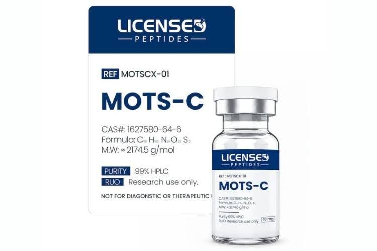
MOTS-c belongs to a small group of peptides called mitochondrial-derived peptides (MDPs), which are made by mitochondrial DNA. It has become a popular area of interest in current cellular biology since the peptide was discovered in 2015. Researchers are interested in how it communicates with cellular energy regulation pathways and stress responses, as well as metabolic homeostasis, balancing how cells make, use, and store energy.
Interest in MOT-c benefits has expanded because laboratory evidence has shown that it may affect several biological processes, such as glucose metabolism, mitochondrial signaling, and cellular energy sensing. As a result, scientists have been able to better understand how these MDPs help with metabolic balance while environmental conditions change, although research is still limited to preclinical settings.
This blog will consider the current literature regarding the MOTS-c peptide’s benefits with a focus on research applications, and we’ll look at the biological origin of MOTS-c, its mechanisms of action, research findings, and considerations.
Please Note: USA-made research peptides are for research use only and not for human consumption, clinical use, or diagnostic purposes.
What Is MOTS-c?

MOTS-c is the common name for Mitochondrial Open Reading Frame of the Twelve S rRNA Type-c, and its distinguishing feature is its genetic origin. It’s a short peptide made up of 16 amino acids. What differentiates if from other peptides involved in intracellular signaling is that MOTS-c is encoded in the mitochondrial genome and not the nuclear DNA.1
Laboratory evidence indicates that MOTS-c might interact with multiple metabolic pathways at the same time, which is why it’s become an important topic in mitochondrial biology research.2
Importantly, the peptide has not been approved for human consumption or clinical applications.
Discovery of Mitochondrial-Derived Peptides
Mitochondria are well known as a cell’s main energy-producing organelles; however, one of the first mitochondrial-derived peptides discovered was Humanin, which, along with the later discovery of Small Humanin-Like Peptides (SHLPs), revealed that mitochondria take part in cellular signaling, not just energy production.2
Lee et al., who discovered MOTS-c, found that it’s encoded with the 12S ribosomal RNA (rRNA) region of mitochondrial DNA, revealing that previously overlooked areas of the DNA found inside the mitochondria can actually make functional peptides.7
Most mitochondrial proteins stay inside the mitochondria, but preclinical studies show that during metabolic stress, MOTS-c can travel to other parts of the cell. What’s being investigated is how this movement helps cells adapt when their energy demands change.
Primary Cellular Mechanisms of Action
As mentioned, instead of acting via a single receptor, experimental research suggests that MOTS-c is part of multiple signaling networks linked to metabolic adaptation.
These three mechanisms have amassed the most research attention:
- Pathways associated with insulin sensitivity and resistance, and metabolic dysfunction
- AMP-activated protein kinase (AMPK) signaling
- Glucose metabolism and uptake
Activation of AMPK and Energy Regulation
AMPK is a primary energy sensor in the cell. When it detects that energy availability is decreasing, AMPK helps to restore metabolic homeostasis, or balance, by regulating the pathways that manage how the body uses nutrients, keeps mitochondria working, and helps cells adapt to stress and changing energy levels.3
Preclinical research shows that when under induced metabolic stress (experimentally), MOTS-c might trigger AMPK signaling. After animal models and cultured cells were exposed to the peptide, researchers noticed increased activity in the AMPK-linked pathways.4
These findings have made AMPK one of the most-studied mechanisms in terms of MOTS-c benefits.
Regulation of Glucose Metabolism and Uptake
Glucose metabolism is another area of focus. Because glucose supplies a principal source of cellular energy, efficient glucose uptake is crucial for regular metabolic function.
Laboratory-based research indicates that MOTS-c may help manage how cells take in and use glucose for energy; however, rather than acting as a glucose transporter itself and moving the glucose into the cells, it seems to work with the cellular signaling networks, e.g., the AMPK pathway.5
These findings are helping to enhance scientific understanding of how glucose metabolism and uptake, and mitochondrial signaling, engage.
Notably, all evidence to date comes from preclinical studies and not human research.
Influence on Insulin Sensitivity Pathways
Another area that researchers are investigating is how MOTS-c engages with the pathways connected to insulin sensitivity and resistance, but with a focus on molecular signaling, not disease outcomes. Experimental studies show that it may be part of the signaling networks associated with nutrient sensing and regulating cellular energy in metabolic-dysfunction conditions.7
The laboratory research evidence shows that MOTS-c works through several interconnected pathways, instead of a singular molecular target, which is an indication of the broader role mitochondria play in cellular communication.2
Metabolic Pathways Observed in Preclinical Research
The main areas of investigation are:
- Mitochondrial Biogenesis: Developing new cells from pre-existing cells to meet energy demands
- Lipid Metabolism: The processing of fat
- Cellular Adaptations Seen in Exercise-Mimetic Models: Cell changes that imitate exercise
Mitochondrial Biogenesis and Cellular Respiration
Mitochondrial function is important when it comes to regulating metabolism and producing cellular energy, and research suggests that MOTS-c might influence the signaling pathways associated with mitochondrial biogenesis, including those shaped by AMPK and PGC-1α, a protein that helps cells make more mitochondria. MOTS-c doesn’t act directly on the mitochondria; however, it appears to be part of signaling networks that help cells adapt to changing energy needs.5

[Photo by Nuchao, Canva].
Lipid Metabolism in Lab Models
The next area of research regarding MOTS-c is its connection to lipid metabolism. Scientists have found evidence suggesting it may affect the signaling pathways that help cells use fatty acids as an energy source during metabolic stress. However, the findings are based solely on preclinical lab research.7
The interest is mostly around how MOTS-c potentially affects fat transport and oxidation, and energy use in cells in general. There is also some research exploring if it acts on metabolic flexibility, which is a cell’s ability to switch between different energy sources. This metabolic flexibility helps cells adapt when the availability of nutrients changes.7
Cellular Synergy in Exercise-Mimetic Research
The next primary area of research regarding MOTS-c is in exercise-mimetic lab models. Essentially, MOTS-c has been found to activate several overlapping exercise-related cellular pathways in controlled lab studies.8,9
Researchers also noted changes in how cells turn genes on and off, otherwise known as gene expression, when animal models and cultured cells were exposed to MOTS-c.9
Importantly, researchers use the term exercise-mimetic because this peptide appears to activate some of the same cellular signaling pathways as exercise does, but that doesn’t mean that MOTS-c can produce the same effects as regular exercise or replace physical activity.8,9
What Have Animal and Cell Studies Actually Observed?
Most of what is known about MOTS-c comes from a handful of well-cited rodent and cell-culture experiments. The findings below are laboratory observations in models, not outcomes in people, and none of them establish a clinical effect.
Physical Performance in Aged Rodents
In one of the most-cited experiments in this area, young, middle-aged and old mice were given MOTS-c and then assessed on rotating-rod balance tests and progressively faster treadmill runs. Treated animals in every age group ran longer and produced more work than untreated controls, and a proportion of the old treated mice completed a final high-speed treadmill stage that no untreated old animal reached.17
The same work reported improvements in grip strength, stride length and timed walking in very old animals, and noted that a late-life, intermittent schedule was enough to produce them — a point researchers highlight because interventions started late in life are easier to model than lifelong ones.17
Cellular Stress Resistance in Muscle Cell Lines
In C2C12 mouse myoblasts, MOTS-c treatment roughly doubled cell survival through 48 hours of glucose restriction and serum deprivation, and treated cells recovered far more strongly after a week of the same stress. When glucose was removed entirely and only lipids were available as fuel, most untreated cells did not survive, while treated cultures again showed about a two-fold improvement. Metabolic flux measurement in the same experiments indicated a greater capacity to use lipids for energy.17
Silencing heat shock factor 1 (HSF1) removed this protection, which identifies HSF1 as one mediator of the peptide’s stress-resistance activity in these cells.17
Plasma Metabolite and Insulin-Sensitivity Measurements
An untargeted metabolomics study in diet-induced obese mice reported that sphingolipid, monoacylglycerol and dicarboxylate pathways — routes typically elevated in obesity models — were downregulated following MOTS-c treatment, alongside lower plasma sphingosine-1-phosphate and reduced fat accumulation in skeletal muscle.18
Separately, hyperinsulinemic-euglycemic clamp work reported roughly a 30% improvement in systemic insulin sensitivity after short-term administration, and the effect was observed independently of any change in body weight in standard-diet animals.7
Cardiac and Vascular Models
In a 12-week treadmill-training study in rats, animals that trained while receiving MOTS-c showed a lower resting heart rate and a higher ejection fraction than animals that trained alone, although most measures of cardiac geometry did not differ between the two trained groups. Myocardial MOTS-c content rose in both, and exogenous administration raised it further; this was accompanied by increased AMPK phosphorylation with no change in total AMPK protein, indicating activation of the existing pool rather than more of it.19
Reviews of the vascular literature describe MOTS-c limiting calcium phosphate deposition and disorganised elastic fibres in models of induced vascular calcification, an effect linked to restoring AMPK activity that the model had suppressed.9 Low circulating MOTS-c has also been reported in people with coronary endothelial dysfunction, which is an association measured in patients, not evidence of an effect.21
Osteogenic Models
Work in bone marrow mesenchymal stem cells reported that MOTS-c increased calcified nodule formation and the expression of genes in the TGF-β/Smad pathway, pushing those cells toward an osteoblast phenotype; inhibiting TGF-β1 removed the effect.22 This is a single cell-model finding and remains a minor branch of the literature compared with the metabolic work.
Do MOTS-c Levels Change With Age and Exercise?
Human sampling studies have measured MOTS-c rather than administered it, and the pattern is not uniform across tissues.
In healthy young men performing stationary cycling, skeletal-muscle MOTS-c rose sharply during exercise and stayed partially elevated after a recovery period, while plasma levels rose more modestly and returned to baseline.17 This is the basis for describing MOTS-c as exercise-induced.
Across age groups the picture differs by compartment: circulating levels tend to be lower in older men than in young adults, while skeletal-muscle expression is higher. That difference tracks with muscle fibre composition — MOTS-c expression correlates positively with slow-fibre markers (MYH7) and negatively with fast-fibre markers (MYH2), and ageing muscle shifts toward the slow, mitochondria-dense fibre type.20
Population genetics adds a further thread: a variant in the MOTS-c coding region has been reported at higher frequency in long-lived Japanese cohorts, which researchers cite as a reason to study the peptide in the context of ageing biology.10
How Does MOTS-c Interact With Inflammatory Signaling?
Mitochondrial-derived peptides are sometimes grouped with other mitokines as candidate regulators of the low-grade inflammatory state associated with ageing. In cell and animal models, MOTS-c has been reported to lower pro-inflammatory cytokine output (IL-6, IL-1β, TNF-α), raise the anti-inflammatory cytokine IL-10, and dampen NF-κB and MAPK signalling while promoting STAT3 and aryl hydrocarbon receptor activity.2,9
In severe bacterial infection models, treated animals showed lower bacterial loads and a shifted cytokine profile alongside greater macrophage bactericidal activity.9 These remain model observations; MOTS-c has not been evaluated as a treatment for infection or inflammation in humans.
What Is the Connection to Cellular Senescence?
Senescent cells stop dividing but keep secreting a mix of cytokines, chemokines and matrix-remodelling enzymes known as the senescence-associated secretory phenotype (SASP). Kim and colleagues reported that mitochondrial peptides including MOTS-c modulate mitochondrial function in senescent cells and alter SASP output.1
This has drawn interest because senolytic compounds such as FOXO4-DRI, which clears senescent cells by disrupting the p53–FOXO4 interaction, appear more potent when SASP signalling is high and less potent when it is suppressed.23 The open question researchers are testing is whether raising SASP output makes senescent cells easier for the immune system and for senolytics to identify. It is an early hypothesis built on separate lines of evidence, not a demonstrated combination effect.
Comparing the Structure of Mitochondrial-Derived Peptides
MOTS-c is part of a family of mitochondrial-derived peptides, encoded in mitochondrial DNA, that have the same origin but are different when it comes to biological activity, structure, and placement. By comparing the different peptides, research teams can better understand their various signaling roles.
Structural Differences: MOTS-c vs. Humanin
Humanin is made up of 24 amino acids and is encoded in a different part of the mitochondrial genome, whereas MOTS-c, a shorter peptide, is made up of 16 amino acids and is encoded in the 12S rRNA region.10
When it comes to functional differences, research findings suggest that humanin mostly engages with the stress-response signaling pathways, while MOTS-c seems to be more linked to energy sensing and metabolism regulation.10
Targeted Cellular Pathways in Laboratory Analysis
Current research mainly focuses on the following pathways:
- Insulin sensitivity
- Metabolic homeostasis
- Mitochondrial function
- Glucose metabolism
- Glucose uptake
- Nuclear gene regulation
- Cellular stress adaptation
The following techniques are used in lab models to study how the MOTS-c peptide engages with cellular signaling:11
- RNA sequencing (measuring which genes are active)
- Quantitative PCR (a gene-measurement test)
- Proteomics (analyzing proteins in cells)
- Metabolic profiling (tracking how cells use energy)

[Photo by Getty Images, Canva].
What Makes a Research-Grade Peptide Usable?
Small differences in purity or contamination change experimental results, so material quality is part of the experiment rather than a detail around it. Research teams generally look for third-party analytical testing and batch-level documentation before a compound goes into a protocol.
Every Licensed Peptides batch is verified to a 99%+ purity standard and ships with a batch-specific Certificate of Analysis confirming purity, identity and quality, with testing performed by ISO/IEC 17025-accredited independent laboratories. Batches are also screened for contaminants that can distort results — we publish endotoxin reports and run sterility and heavy metal testing as part of quality control. For the underlying methods, see our guide on achieving and verifying peptide purity.
Where Is Mitochondrial Peptide Research Heading?
The open question in this field is how mitochondrial-derived peptides coordinate signalling between the mitochondria and the nucleus as metabolic conditions change.12 Advances in transcriptomics, metabolomics, proteomics and single-cell sequencing are letting research teams map those networks at a resolution that was not available when MOTS-c was first described in 2015.11 As those tools improve, more of the biology encoded in mitochondrial DNA is likely to be characterised.11,12
How Should MOTS-c Be Stored and Handled in the Lab?
Peptide stability depends on storage and handling. Heat, moisture, prolonged light exposure and repeated freeze-thaw cycles all drive degradation, so lyophilized material is normally kept frozen or refrigerated per the manufacturer’s instructions and reconstituted only as needed.13 Temperature control, correct labelling and careful sample tracking are what keep results attributable to the experiment rather than to a change in the material.13

[Photo by Prathan Chorruangsak, Canva].
FAQs
Is MOTS-c Approved for Human Consumption?
MOTS-c is not approved for human consumption; research is currently at the laboratory and preclinical stage of investigation. At Licensed Peptides, we supply MOTS-c as strictly a research-use-only (RUO) material that’s solely intended for analytical applications. It hasn’t been approved for therapeutic, diagnostic, clinical, or medical use.14
Does MOTS-c Raise Testosterone?
Research to date has mainly investigated MOTS-c regarding its role in metabolic signaling, glucose metabolism, and mitochondrial function; there is no validated evidence proving that MOTS-c directly raises testosterone production. 7
Is MOTS-c Prohibited in Competitive Sports?
Yes, MOTS-c is on the World Anti-Doping Agency (WADA) Prohibited List; it falls under the peptide hormones, growth factors, mimetics, and related substances categories. It is prohibited at all times under Section S4.4.1 (Hormone and Metabolic Modulators, as an AMPK activator).15 However, this classification is related to anti-doping rules for competitive sport and shouldn’t be interpreted as evidence of approved human use. 15 Licensed Peptides only supplies MOTS-c for laboratory research use.
Why Is MOTS-c Called an “Exercise Mimetic”?
Because exercise raises MOTS-c levels in skeletal muscle, and because giving the peptide to animals activates several of the same signalling pathways that exercise does.17 The label describes an overlap in cellular signalling. It does not mean the peptide reproduces the effects of exercise, and no study has shown that it can substitute for physical activity.
Has MOTS-c Been Studied in Humans?
Only observationally. Published human work has measured MOTS-c in muscle biopsies and blood samples — before and after exercise, across age groups, and in patients with endothelial dysfunction.17,20,21 There are no published human trials in which MOTS-c was administered, which is why every mechanism described above is reported from cell cultures and animal models.
How Do Researchers Verify Peptide Purity and COAs?
To ensure a peptide is of suitable quality, researchers will check the batch’s Certificate of Analysis (COA), using two primary tests in a lab:16
- HPLC Testing: This measures the overall purity through separation of the main peptide from impurities. The aim is to be left with a single peak covering 99% of the sample.
- Mass Spectrometry: This measures the weight of the molecules to confirm whether it is the correct peptide and not an alternative compound.
They’ll also examine the COA to make sure the batch doesn’t contain any dangerous contaminants that could ruin a lab experiment, such as heavy metals, bacterial endotoxins, and microbes. Lastly, they’ll double-check that the lot number on the physical vial matches the independent test results on the COA.16
Conclusion: MOTS-c Benefits
Interest in the MOTS-c peptide has grown substantially since it was first discovered in 2015. Even though the research is still in the preclinical stage, researchers are continuing to improve understanding of how this mitochondrial-derived peptide participates in regulating energy and communication between cells.
The current evidence we have is mostly derived from in vitro studies using cultured cells and in vivo animal models, and the investigations are examining the molecular mechanisms rather than the clinical outcomes.
But as mitochondrial biology continues to progress, scientists are exploring how these structures communicate with cells and adapt to changing metabolic demands, uncovering new questions that may influence future investigations.
References
- Kim SJ, Mehta HH, Wan J, et al. Mitochondrial peptides modulate mitochondrial function during cellular senescence. Aging (Albany NY). 2018;10(6):1239-1256.
- Zheng Y, Wei Z, Wang T. MOTS-c: A promising mitochondrial-derived peptide for therapeutic exploitation. Front Endocrinol (Lausanne). 2023;14:1120533.
- Hardie DG, Schaffer BE, Brunet A. AMPK: An energy-sensing pathway with multiple inputs and outputs. Trends Cell Biol. 2016;26(3):190-201.
- Herzig S, Shaw RJ. AMPK: guardian of metabolism and mitochondrial homeostasis. Nat Rev Mol Cell Biol. 2018;19(2):121-135.
- Yang B, Yu Q, Chang B, et al. MOTS-c interacts synergistically with exercise intervention to regulate PGC-1α expression, attenuate insulin resistance and enhance glucose metabolism in mice via AMPK signaling pathway. Biochim Biophys Acta Mol Basis Dis. 2021;1867(6):166126.
- Kim KH, Son JM, Benayoun BA, Lee C. The mitochondrial-encoded peptide MOTS-c translocates to the nucleus to regulate nuclear gene expression in response to metabolic stress. Cell Metab. 2018;28(3):516-524.e7.
- Lee C, Zeng J, Drew BG, et al. The mitochondrial-derived peptide MOTS-c promotes metabolic homeostasis and reduces obesity and insulin resistance. Cell Metab. 2015;21(3):443-454.
- Li S, Wang M, Ma J, et al. MOTS-c and exercise restore cardiac function by activating of NRG1-ErbB signaling in diabetic rats. Front Endocrinol (Lausanne). 2022;13:812032.
- Wan W, Zhang L, Lin Y, et al. Mitochondria-derived peptide MOTS-c: effects and mechanisms related to stress, metabolism and aging. J Transl Med. 2023;21(1):36.
- Kim SJ, Miller B, Kumagai H, Silverstein AR, Flores M, Yen K. Mitochondrial-derived peptides in aging and age-related diseases. GeroScience. 2021;43(3):1113-1121.
- Silverstein AR, Flores MK, Miller B, et al. Mito-Omics and immune function: Applying novel mitochondrial omic techniques to the context of the aging immune system. Transl Med Aging. 2020;4:132-140.
- Kim SJ, Xiao J, Wan J, Cohen P, Yen K. Mitochondrial-derived peptides as novel regulators of metabolism. J Physiol. 2017;595(21):6613-6621.
- Sigma-Aldrich. Handling and Storage Guidelines for Peptides and Proteins. Life Science Technical Documents; 2024.
- U.S. Anti-Doping Agency (USADA). What is the MOTS-c Peptide? USADA Education & Science Resources; 2024.
- World Anti-Doping Agency. The 2026 Prohibited List: International Standard. World Anti-Doping Code. Valid January 1, 2026. Accessed July 31, 2026.
- Verbeken M, D’Hondt M, Wynendaele E, De Spiegeleer B. Quality evaluation of peptide drugs: a focus on impurities and analytical characterization. J Pharm Biomed Anal. 2014;101:103-116.
- Reynolds JC, Lai RW, Woodhead JST, et al. MOTS-c is an exercise-induced mitochondrial-encoded regulator of age-dependent physical decline and muscle homeostasis. Nat Commun. 2021;12:470. doi:10.1038/s41467-020-20790-0
- Kim SJ, Miller B, Mehta HH, et al. The mitochondrial-derived peptide MOTS-c is a regulator of plasma metabolites and enhances insulin sensitivity. Physiol Rep. 2019;7(13):e14171. doi:10.14814/phy2.14171
- Yuan J, Wang M, Pan Y, et al. The mitochondrial signaling peptide MOTS-c improves myocardial performance during exercise training in rats. Sci Rep. 2021;11:20077. doi:10.1038/s41598-021-99568-3
- D’Souza RF, Woodhead JST, Hedges CP, et al. Increased expression of the mitochondrial derived peptide, MOTS-c, in skeletal muscle of healthy aging men is associated with myofiber composition. Aging (Albany NY). 2020;12(6):5244-5258. doi:10.18632/aging.102944
- Qin Q, Delrio S, Wan J, et al. Downregulation of circulating MOTS-c levels in patients with coronary endothelial dysfunction. Int J Cardiol. 2018;254:23-27. doi:10.1016/j.ijcard.2017.12.001
- Hu BT, Chen WZ. MOTS-c improves osteoporosis by promoting osteogenic differentiation of bone marrow mesenchymal stem cells via TGF-β/Smad pathway. Eur Rev Med Pharmacol Sci. 2018;22(21):7156-7163. PMID: 30468456
- Baar MP, Brandt RMC, Putavet DA, et al. Targeted apoptosis of senescent cells restores tissue homeostasis in response to chemotoxicity and aging. Cell. 2017;169(1):132-147.e16. doi:10.1016/j.cell.2017.02.031

